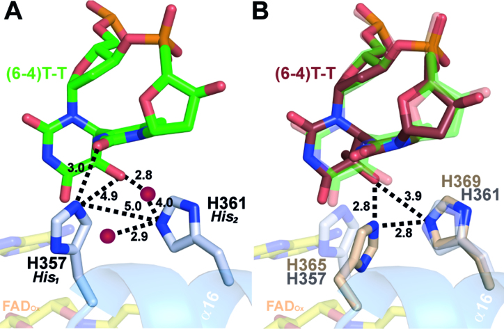
Figure 5.
Conformation of the active site histidines in (6-4) photolyases. (A) Enhanced view of the (6-4)PP interactions with the two prominent residues H357 (His1) and H361 (His2) of CraCRY (gray). Distances (in Å) were measured between the histidines, the DNA lesion and two water atoms (red). (B) Comparison of the active site of CraCRY (6FN0, gray) with Dm(6-4) (3CVU, wheat) (r.m.s.d. 0.658 Å). The (6-4)PP bound to Dm(6-4) (red) is slightly tilted compared to the (6-4)PP bound to CraCRY (green).

