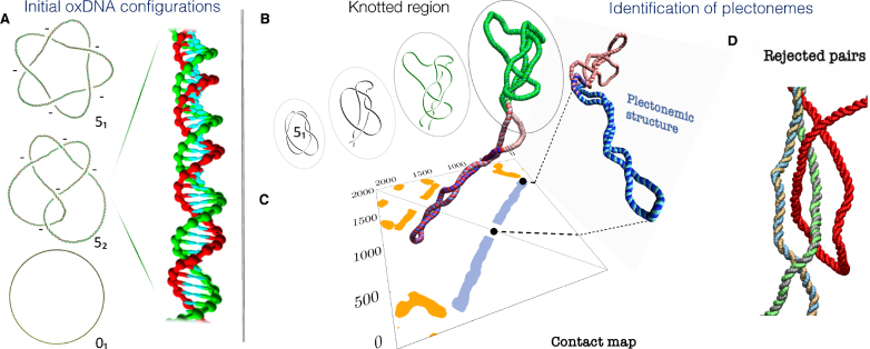
Figure 1.
(A) Initial configurations of the supercoiled double-stranded DNA rings for the three considered topologies, 01, 51 and 52. The latter two are left-handed, i.e. the topological sign of their projected crossings is negative, as indicated. The 51 and 52 snapshots have been edited to highlight the over- and underpasses, see Supplementary Figure S1 for the unedited versions. The mesoscopic structural representation of the oxDNA model is illustrated in the inset, which shows a magnified portion of one of the rings. The twist was uniformly adjusted for each of the three cases to yield the same level of negative supercoiling (-5%). (B–D) Identification of the knotted and the plectonemically-wound regions for a typical 51-knotted supercoiled conformation, shown in the foreground. (B) The knotted region (green) is the shortest portion that, after suitable bridging of the termini, has the same (51) topology of the entire ring. (C) The plectonemically-wound region (blue) is found by using the contact map to identify extended superhelical regions ending in a short apical loop and that are free of cis or trans entanglement, as in the case of panel (D).

