Figure 8.
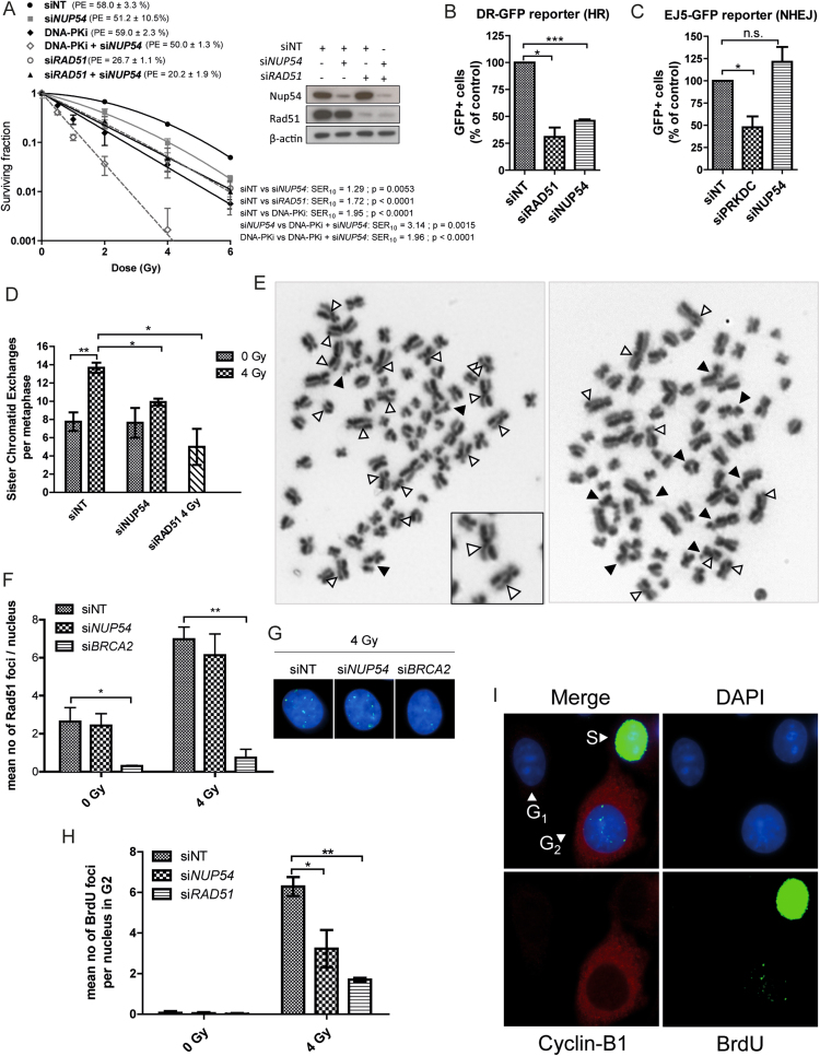
Nup54 depletion does not further radiosensitize HR-deficient cells and impairs HR-mediated repair. (A) CFA of HeLa cells transfected with siNT, siNUP54 and siRAD51, alone or in combination (40 nM siNT (siNT); 20 nM siNT + 20 nM siNUP54 (siNUP54); 20 nM siNT + 20 nM siRAD51 (siRAD51); 20 nM siNUP54 + 20 nM siRAD51 (siRAD51 + siNUP54)). One hour before IR exposure, 1 μM NU7441 (DNA-PK inhibitor (DNA-PKi)) was added to siNT and siNT + siNUP54-treated cells (DNA-PKi and DNA-PKi + siNUP54, respectively), and removed 24 h after IR exposure. Surviving fraction, SER10 and statistical significance of differences between curves were estimated as in Figure 1. (B) HR repair efficiency determined with the DR-GFP reporter in HEK293 treated with the indicated siRNAs. siRAD51 was used as a positive control. (C) NHEJ repair efficiency determined with the EJ5-GFP reporter in HEK293 treated with the indicated siRNAs. An siRNA targeting DNA-PK (siPRKDC) was used as a positive control. (D) Mean number of SCEs per metaphase in HeLa cells accumulated in mitosis from 8 to 12 h after 4 Gy IR. (E) Examples of metaphase spreads of siNT- (right) and siNUP54-treated (left) cells. SCEs and chromatid-type aberrations are indicated with white and black arrows, respectively. Examples of SCEs are shown at a higher magnification (inset). (F) Mean number of Rad51 foci per nucleus in HeLa cells at 6 h after IR. siBRCA2 was used as a positive control for Rad51 foci formation inhibition. (G) Image of Rad51 foci in siNT- and siNUP54-treated cells 6 h after 4 Gy IR (green: Rad51; blue: DAPI). (H) Mean number of BrdU foci detected after DNase treatment in HeLa cells irradiated with 4 Gy in G2 phase (6 h after IR). (I) Staining pattern of cells from experiment depicted in H. With the exception of graph shown in A, values correspond to mean ± sd from three independent experiments (*P < 0.05; **P < 0.005; ***P < 0.001; n.s.: non-significant; two-sided t-test).

