Figure 1.
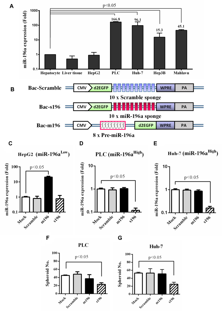
Involvement of miR-196a in HCC tumorigenicity. (A) MiR-196a expression in different HCC cells. (B) Recombinant BVs that expressed scramble sponge (Bac-Scramble), miR-196a sponge (Bac-s196) and pre-miR-196a (Bac-m196). (C–E) miR-196a levels in HepG2 (C), PLC (D) and Huh-7 (E) cells after BV transduction. (F–G) Spheroid formation in PLC (F) and Huh-7 (G) cells after BV transduction. The miR-196a levels in various HCC and normal cells were quantified by TaqMan® RT-qPCR and normalized to that in human hepatocytes. HCC cells were mock-transduced or transduced with recombinant BVs at MOI 200 and the miR-196a levels were analyzed by RT-qPCR at 1 dpt. The data represent means±SD of triplicated culture experiments.

