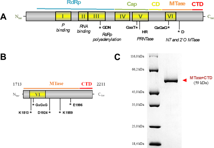
Figure 1.
The MTase+CTD domain of Sudan ebolavirus L protein. (A) Sequence analysis of the mononegavirus L protein revealed six conserved regions (CRI to CRVI, yellow boxes) that contain motifs responsible for the different activities of the L (motifs mapped with asterisks) (Poch et al., 1990). Additionally, the recently published VSV L structure resolved by cryo-EM shows that the L protein is organized in 4 domains: the RNA-dependent RNA polymerase (RdRp) intricated with the polyribonucleotidyltransferase (PRNTase or Cap), a connector domain, the methyltransferase domain (MTase) and a poorly conserved C-terminal domain (CTD). (B) Based on alignments with the VSV L protein, the MTase+CTD domain in SUDV L protein was defined as the fragment covering amino acids 1713–2211. SAM-binding site motifs (GxGxG) and the 2’O catalytic tetrad K-D-K-E have also been identified (asterisks). (C) SDS-PAGE of purified, recombinant SUDV MTase+CTD containing an N-terminal oligohistidine-tag (58.4 kDa).

