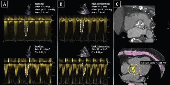
Figure 4: Classical Low-flow, Low-gradient Severe Aortic Stenosis.

A: A 75-year old male with ischaemic cardiomyopathy, reduced left ventricular ejection fraction (32 %) and low cardiac output. At rest, echocardiography showed calcified aortic valve with severely narrowed valve area <1.0 cm2, while peak velocity and mean gradient were in the range of moderate aortic stenosis. B: During low-dose dobutamine stress echocardiography peak jet velocity and mean gradient increased ≥4.0 m/s and ≥40 mmHg respectively and the aortic valve area remained <1.0 cm2, revealing true severe aortic stenosis. Furthermore, an increase in cardiac output demonstrated left ventricular contractile reserve. C: CT showed a tricuspid aortic valve with high calcium score, suggesting high likelihood of severe aortic stenosis. AU = arbitrary units; AVA = aortic valve area; CI = cardiac index; Mean gr = mean gradient; SVi = stroke volume index; Vmax = peak velocity.
