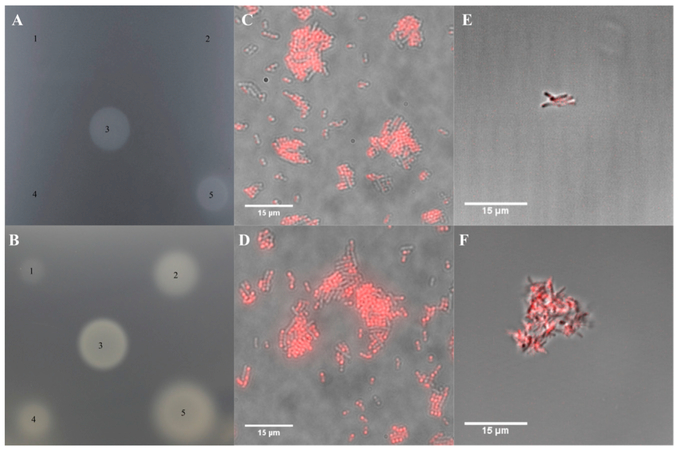
Figure 5. Effect of Cobyric Acid and Cobalamin Analogs on Bacteria.
(A) A bioassay plate containing an S. enterics strain that requires cobyric acid or later pathway intermediates for growth. Addition of a known amount of vitamin B12 (spot 3) allows growth. Spots 1, 2, and 4 represent C5-allyl-hydrogenobyrinic acid a,c-diamide (C5-allyl-HBAD), C5-allyl-Hby, and C5-(TPEA)-Hby, none of which promote growth. Spot 5 contains the C5-(TPEA)-cobyric acid analog and promotes growth.
(B) The same bioassay plate assay as in (A), with spot 3 containing a standard vitamin B12 solution. Spots 1, 2, 4, and 5 are solutions of C5-BoD-co-byricacid, ribose-BoD-cobalamin, C5-OG-cobyric acid, and ribose-OG-cobalamin, respectively. All the analogs support growth although the C5 analogs are weaker than the respective ribose-linked analogs.
(C and D) Overlay pictures of E. coli cells that have been grown in the presence of ribose-BoD-cobalamin and C5-BoD-cobyric acid, respectively. The co-localization of the red fluorescence with the cells indicates that E. coli has taken up both compounds.
(E and F) Cultures of M. tuberculosis, grown in the presence of ribose-BoD-cobalamin and C5-BoD-cobyric acid, respectively. Again, the presence of the red fluorescence with the cells indicates that M. tuberculosis is able to take up both analogs.

