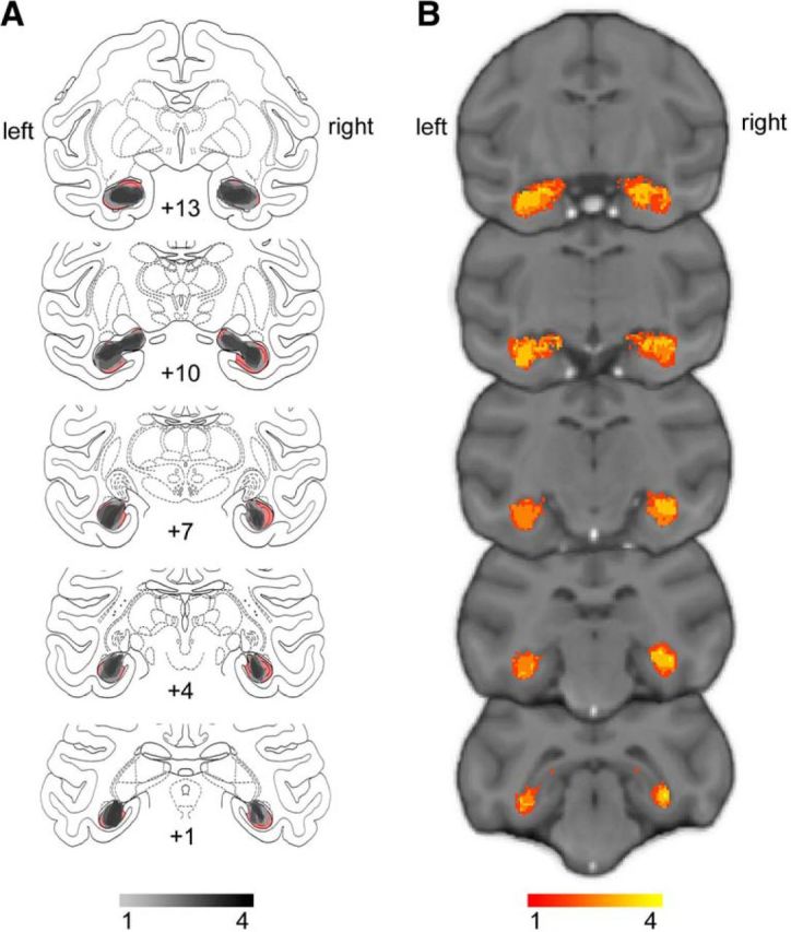
Figure 2.

Extent and variability of hippocampal lesions. A, Sketch of hippocampal size based on histology (Nissl-stained sections) overlaid on atlas sections. The unlesioned hippocampal volume is shown in red. Overlap of remaining hippocampal volume is shown for the four monkeys indicating shrinkage of the hippocampus bilaterally in all monkeys. B, T2-weighted hypersignal 6 d after surgery indicating local inflammation in the hippocampus. Overlap is shown for the four monkeys.
