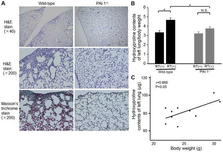
Figure 5.
(A) Histological analyses using hematoxylin and eosin or Masson's trichrome staining in sections of irradiated lung at 24 weeks after irradiation. Scale bar=100 µm. (B) Hydroxyproline contents of the left lung per body weight at 24 weeks after irradiation. Data are shown as mean ± SEM for 5–6 mice per group. *P<0.05 as indicated. (C) Correlation between hydroxyproline contents of the left lung and body weight in mice (n=10). PAI-1, plasminogen activator inhibitor-1; H&E, hematoxylin and eosin.

