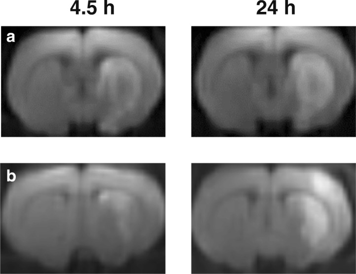
Fig. 1.

Characterization of ischemia using magnetic resonance imaging. A typical diffusion-weighted imaging (DWI) of the formation of ischemic injury areas with a subcortical localization both 4.5 and 24 h after tMCAO (a), as well as subcortical and hemispheric localization at 4.5 and 24 h after tMCAO, respectively (b) in the rat brain
