Figure 1.
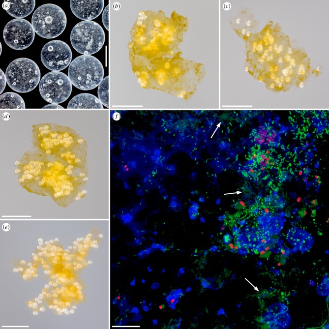
Microplastics and experimentally formed aggregates consisting of biogenic particles and microplastics. (a) Stereo micrograph showing some of the polystyrene beads used for the experiments. (b–e) Photographs of exemplary aggregates that formed out of biogenic particles and microplastics during the experiments with clean microplastics (b) and with biofilm-covered microplastics (c–e). (f) Confocal laser scanning micrograph exhibiting a biofilm formed within five weeks on the surface of one of the polystyrene beads used for the experiments. Blue, polysaccharide-containing structures; green, structures containing nucleic acids; red, chlorophyll-containing structures. The arrows indicate exemplary structures containing extracellular DNA. Scale bars: 500 µm (a), 5 mm (b–e) and 20 µm (f).

