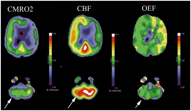
Figure 3.
Diaschisis: This patient has a complete atherosclerotic occlusion of the right internal carotid artery and previous stroke in this territory. Consequently, the CMRO2 image demonstrates reduced oxygen metabolism relative to the contralateral hemisphere (white arrow). The reduced metabolic activity in the right frontal area has caused reduced metabolic activity in the structurally normal left cerebellar hemisphere. This phenomenon is known as diaschisis. The primary reduction in metabolism in the cerebellum leads to a reduction in CBF in both the frontal lobe and the cerebellum (white arrows).

