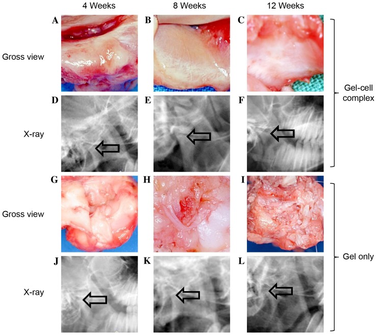
Figure 3.
Monitor repaired defects post-surgery by gross view and x-ray. In gel-cell complex implantation group: (A) At week 4, the surfaces were slightly rough and depressed in gross view, and no Pluronic F-127 gel was found on the defect areas (a); (B) at week 8, the surfaces were smooth and a cartilage-like layer can be observed on the surface; (C) at week 12, the surfaces were smooth and a cartilage-like layer can be observed on the surface. (D-F) The X-ray films showed that there was no obvious bone destruction and no osteophyte was formed. In the gel only group: (G) The surfaces were slightly rough and depressed, and no Pluronic F-127 gel was found; (H) the surfaces were rough and depressed; (I) the surfaces were rough and had some fibrous-like tissues on the surface. (J-L) The X-ray films showed that there was no obvious bone destruction and no osteophyte was formed. In the gel only group, the joint space seemed broadened compared with the gel-cell group (indicated by the arrow, phase contrast, ×100).

