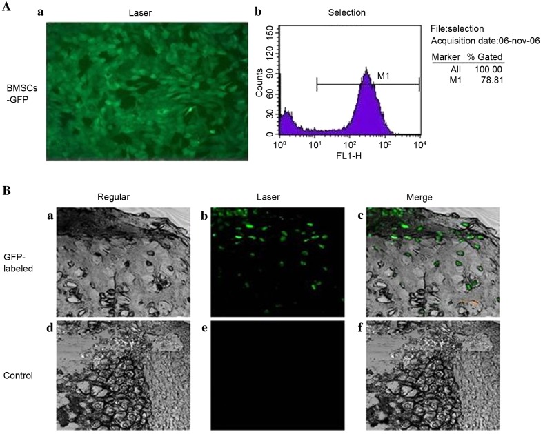
Figure 5.
Evaluation of cartilage repairby confocal microscopy. (A) Quanfication of GFP expression in BMSCs. (a) GFP expression distribution of BMSCs in fluorescent microscopy (×100). (b) GFP-expressed BMSCs selected by flow cytometry. (B) Repaired cartilage comparison between gel-cell complex implantation and control groups were demonstrated by (a and b) confocal microscopy (×200, (c and d) bright field images and (e and f) merged images. GFP-expressed BMSCs were evenly distributed in repaired cartilage area and formed typical lacuna structures at 12 weeks post-surgery. GFP, green fluorescent protein; BMSCs, bone marrow stromal cells.

