Fig. 1.
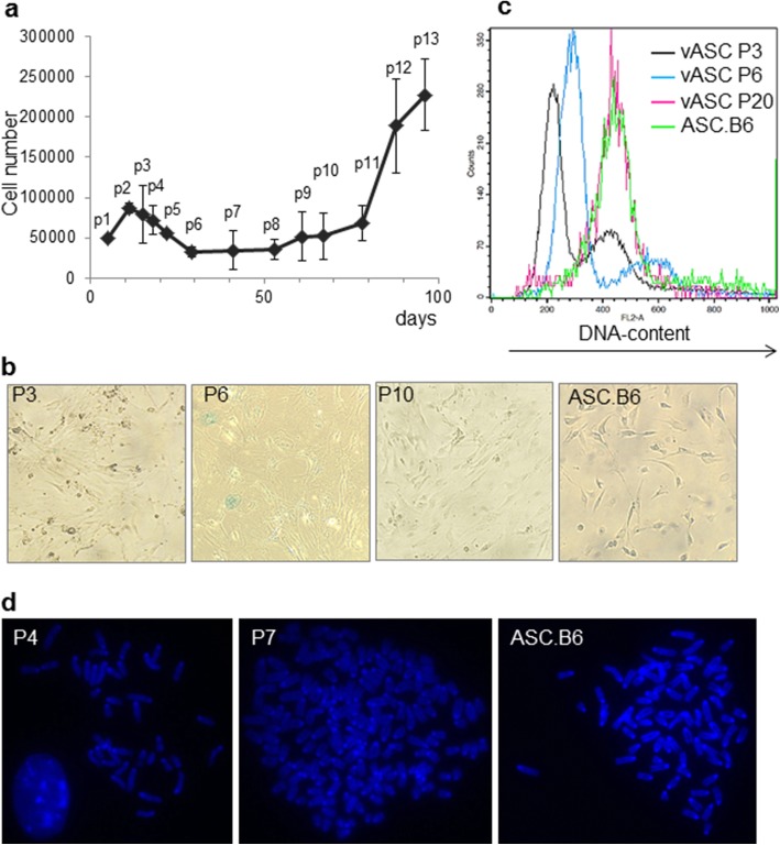
Changes in the proliferation, morphology and ploidy of vASCs under prolonged in vitro culturing. a Fifty thousand vASCs were plated and cultured in triplicate samples. Cells were passaged when the culture reached confluency and the living cell number of vASCs was determined with trypan blue staining and counting with BioRad TC10 counter device. The graph shows the average ± SD of living cell numbers in three parallel samples. The x-axis indicates days in culture from the initial plating, and the measuring points are referred as p1 to p13. A representative of 3 independent experiments is shown. b vASCs at passage numbers 3, 6 and 10 and ASC.B6 cell line was stained for SA-βgal activity for 16 h and then the blue staining was detected with inverted light microscope. c The DNA-content of vASCs at passage numbers 3, 6 and 10 and of ASC.B6 cell line was determined by propidium-iodide staining and flow cytometric analysis. d Metaphase chromosome spreads were made from colchicine-blocked vASCs at passage numbers 4 and 7, and ASC.B6. Chromosomes were DAPI stained and counted using fluorescent microscope

