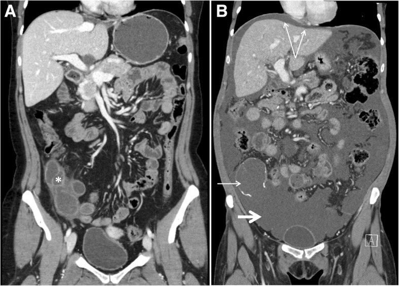
Fig. 1.

Development of pseudomyxoma peritonei (PMP). CT scans with coronal view before (a) and after (b) development of PMP. Mucinous cystadenoma of the appendix (asterisk) in a. Typical features of PMP (b): ruptured calcified appendix (short slim arrow), massive mucinous ascites (short thick arrow), central displacement of the small bowel, and visceral scalloping of the liver (double arrow)
