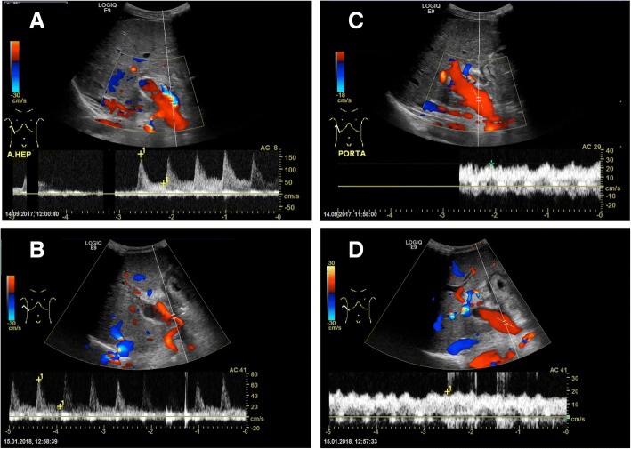
Fig. 2.
Circulation of the liver graft. The pictures to the left show examination of the common hepatic artery of the liver graft with US Doppler before (a) and after (b) CRS-HIPEC with a resistive index (RI) of 0.73 and 0.72 respectively. The pictures to the right show US Doppler examination of the portal vein of the liver graft before (c) and after (d) CRS-HIPEC with no resistance found in either exam

