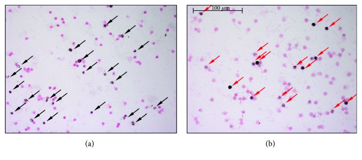
Figure 1.
Immunostaining of leukocyte smears. (a) Representative microscopic image of leukocyte smears stained against nitrotyrosine. Black-colored precipitate indicates the diffuse labeling. Magenta-colored NFR served as counterstain. Black-colored arrows show positively stained cells. (b) Photograph of smear immunolabeled with anti-PAR antibody. Black color represents positive nuclear staining. Counterstaining was NFR. Red arrows point on positive leukocytes.

