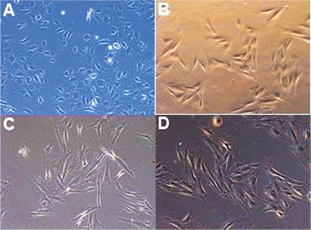
Figure 1. Comparison of phase-contrast microscopic appearance of 3 anti-vascular endothelial growth factor drugs and control culture showed no morphological changes of the retinal pigment epithelium (RPE) cell culture with any drug, and RPE cells maintain the hexagonal morphology at the end of 72 hours in the (A) control, (B) aflibercept (0.5 mg/mL), (C) bevacizumab (0.3125 mg/mL), and (D) ranibizumab (0.125 mg/mL) groups.

