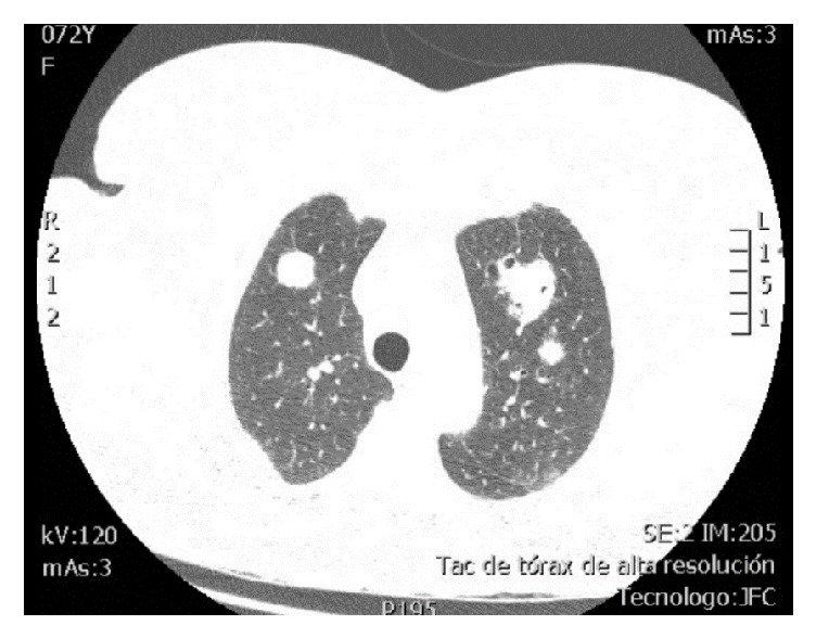
Figure 2.

HRCT of the thorax, axial view, pulmonary window. There are multiple nodules with random distribution in both lung fields, some of which demonstrate discrete halo sign in ground-glass opacity and others demonstrate cavitation. The largest size is subpleural, in the posterior segment of the right upper lobe, measuring 23 x 32 x 30 mm.
