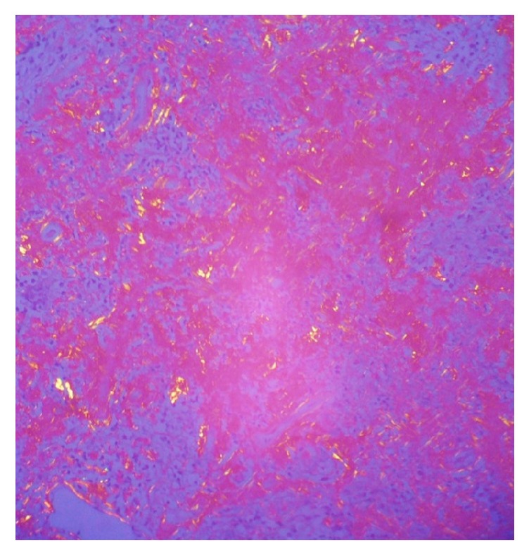
Figure 4.

Hematoxylin and eosin stain in lung biopsy. 4X. Pulmonary parenchyma with distortion of its architecture, anthracite pigment is observed, and alveolar septa thickened with inflammatory infiltrate of lymphocyte predominance. Presence of peribronchiolar lymphocytic inflammatory infiltrate in some lymphoid nodules.
