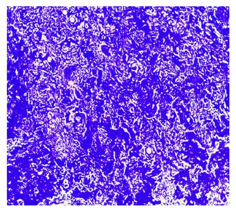
Figure 5.

Crystal violet staining in lung biopsy. 4X. Pulmonary parenchyma where nodules are made up of dense amorphous eosinophilic material that captures the coloration, which confirms the presence of amyloid. In the middle of it, there are inflammatory cells of the lymphoplasmacytic type.
