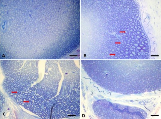
Figure 3.

Light microscopic images of the sciatic nerve from normal control rats (A) and DN rats untreated (B) or treated daily either with 30 mg/kg (C) or 100 mg/kg apocynin (D) for 4 weeks.
Toluidine blue staining was performed. Original magnification 40×, scale bars: 125 µm (A). Red arrows represent degenerated axonal structures (B). Toluidine staining of sciatic nerve from DN rats revealed marked vasoconstruction, increased endothelial cell activation and proliferation, thickening in basal lamina in the capillary vessels (B). There was an increase in extracellular matrix, atrophy in axonal structures, myelin degeneration with loss of Schwann cells and myelin and impairment in configuration (B). Apocynin treatment at both doses reversed these histopathological changes dramatically and dose-dependently prevented Schwann cell degeneration (C and D). DN: Diabetic neuropathy.
