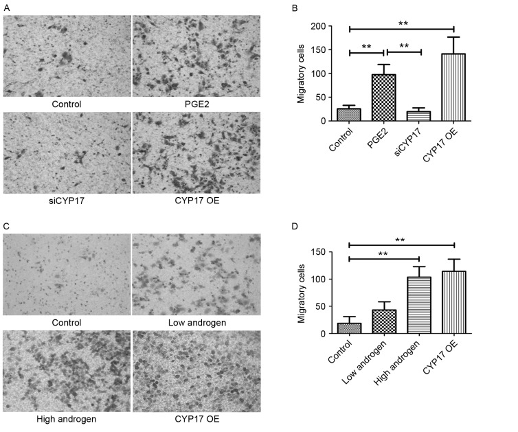
Figure 4.
High expression of CYP17 promotes invasion in endometrial cancer cells. (A) Transwell migration assay images of Ishikawa cells in control, PGE2-stimulated, siCYP17 and CYP17 OE groups. Cells were stained with crystal violet. (B) The number of Ishikawa cells in control, PGE2 stimulated, siCYP17 and CYP17 OE groups (averaged across 5 random images). (C) Transwell migration assay images of Ishikawa cells in control, low androgen (treated with 10−7 g/l androgen for 48 h), high androgen (treated with 10−5 g/l androgen for 48 h) and CYP17 OE groups. Cells were stained with crystal violet. (D) The number of Ishikawa cells in control, low androgen, high androgen and CYP17 OE groups (averaged across five random images). **P<0.01, analyzed by one-way analysis of variance. Micrographs were taken at ×200 magnification. CYP17, cytochrome P450 17α hydroxylase; PGE2, prostaglandin E2; siCYP17, CYP17 small interfering RNA; CYP17 OE, CYP17 overexpression.

