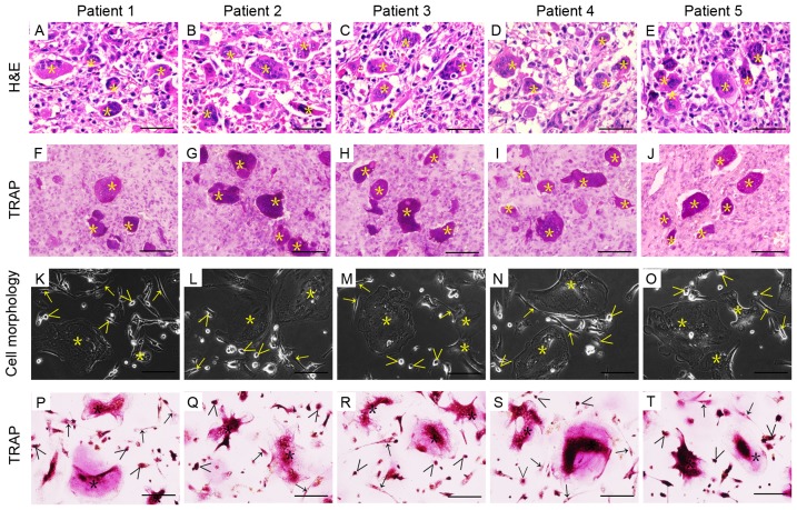
Figure 1.
Histological features of GCTB tissues and their corresponding cell morphology. (A-E) H&E staining of patient tumor tissues with GCTB. (F-J) TRAP-enzyme staining of GCTB tissues. (K-O) Phase-contrast photomicrographs of primary GCTB cells isolated from fresh tumor samples taken from different patients. (P-T) TRAP-enzyme staining revealed TRAP-positive multinucleated osteoclasts in GCTB cells from different patients. Giant cells are marked with asterisks (Õ), monocytes with greater-than signs (>), and stromal cells with arrows (→). Bars represent 50 µm. H&E, hematoxylin and eosin; TRAP, tartrate-resistant acid phosphatase; GCTB, giant cell tumor of bone.

