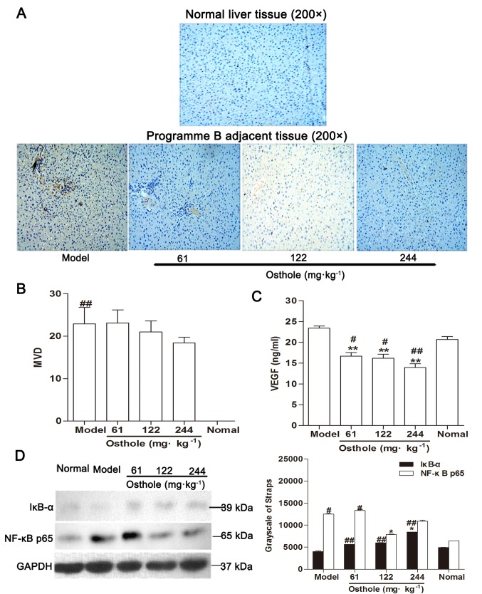
Figure 4.
Effects of osthole on MVD, VEGF and NF-κB signaling in adjacent tissues of orthotopic hepatocellular carcinoma-bearing mice. (A) Immunohistochemical staining for blood vessels with CD34 (arrow) was performed on adjacent tissues from osthole-treated (61, 122 and 244 mg/kg) mice and normal liver tissue sections. (B) Compared with the model control group, the staining indicated that the MVD was decreased in the osthole-treated mice. (C) Expression levels of VEGF in adjacent tissues were measured by ELISA. (D) Adjacent tissue lysates were prepared and quantified. Protein expression of IκB-α and NF-κB p65 were detected by western blot analysis. Equal loading was confirmed by stripping immunoblots and reprobing for GAPDH. Statistical analysis of IκB-α and NF-κB p65 quantification. Data are presented as the mean ± standard error of the mean. *P<0.05, **P<0.01, compared with the model control group; #P<0.05, ##P<0.01, compared with the normal control group. MVD, microvessel density; VEGF, vascular endothelial growth factor; NF-κB, nuclear factor-κB; IκB-α, inhibitor of κB-α.

