Figure 6.
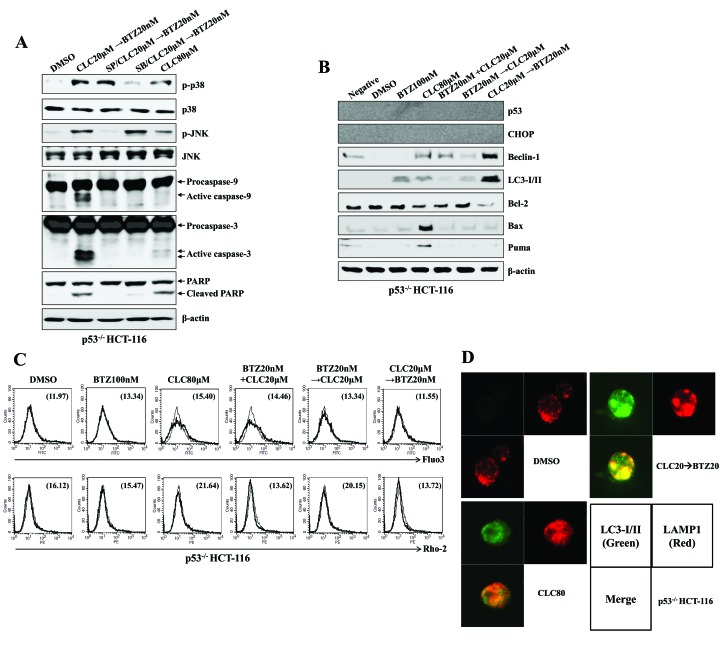
Sequential treatment with CLC and BTZ induces the autophagy-associated cell death of p53−/− colon cancer cells. p53−/− HCT-116 cells were treated with BTZ or CLC alone or in combination. (A and B) Total cell lysates of each condition were harvested and blotted with the indicated antibodies. β-actin served as an internal control. (C) Cytosolic and mitochondrial Ca2+ levels in HCT-116 cells were determined using Fluo3-AM and Rhod2-AM, respectively. (D) Subcellular distribution of LC3-I/II and LAMP1 in p53−/− HCT-116 cells. Cells were observed under a confocal microscope (magnification, ×400). Green fluorescence indicates LC3-I/II, and red fluorescence indicates LAMP1. The results are representative of three independent experiments. CLC, celecoxib; BTZ, bortezomib; LC3-I/II, microtubule-associated protein 1A/1B-light chain 3; LAMP1, lysosomal-associated membrane protein 1; DMSO, dimethyl sulfoxide control; p-, phosphorylated; JNK, c-Jun N-terminal kinase; PARP, poly (ADP-ribose) polymerase; negative, non-treated cells; CHOP, CCAAT-enhancer-binding protein homologous protein; Bax; Bcl-associated X; PUMA, p53-upregulated modulator of apoptosis.

