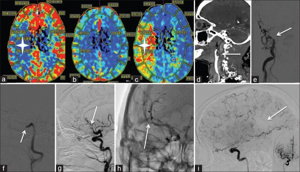
Figure 1.
Wake up Stroke: (a-c) represents perfusion imaging on a 75-year-old patient presenting with a wakeup stroke and NIH Stroke Scale 17. (a) The white star is in the area of low cerebral blood flow. (b) Preserved cerebral blood volume is noted. (c) The white star is in the ischemic penumbra as demonstrated by elevated mean time to transit. (d) A right internal carotid artery cervical occlusion extending to the supraclinoidal internal carotid artery is noted on this sagittal computed tomography angiography (white arrow). (e) Anteroposterior right common carotid artery roadmap with internal carotid artery occlusion at the takeoff (white arrow). (f) Lateral right common carotid artery roadmap. A large bore aspiration catheter was utilized to aspirate the clot to the level of the cavernous internal carotid artery (white arrow). (g) Lateral right internal carotid artery subtracted run revealing an M2 cutoff after recanalization of the internal carotid artery with 1 pass of a 6 mm × 30 mm stentriever (white arrow). (h). Anteroposterior native right internal carotid artery run with a 4 mm × 40 mm stentriever in the M2 (white arrow). (i) thrombolysis in cerebral infarction 2b recanalization with a medium size parietal perfusion deficit (white arrow). Inset is the anteroposterior view right internal carotid artery postthrombectomy. 24 h postprocedure the patient was NIH Stroke Scale 3

