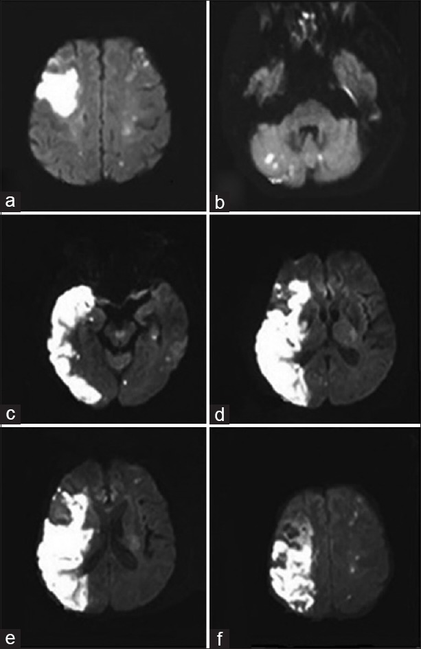
Figure 1.

(a-b) Diffusion weighted imaging showed multiple lesions in bihemispheric territories and cerebellum (2016/2/4) (c-f) Diffusion weighted imaging showed massive ischemic infarction in the right cerebral hemisphere as well as multiple punctate infarcts in the brainstem, bilateral cerebellum and left cerebral hemisphere (2016/2/15)
