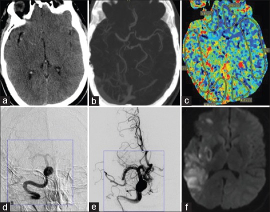
Figure 1.

A 50-year-old male with a history of hypertension woke up with difficulty walking and left side weakness with a summated NIH stroke scale of 7. (a) Initial noncontrast computed tomography demonstrated hyperdense right middle cerebral artery vessel and early ischemic changes. (b) Computed tomography angiogram demonstrated mid-distal right M1 segment occlusion. (c) Computed tomography perfusion shows perfusion mismatch in right middle cerebral artery territory. (d and e) Digital subtraction angiogram showing right M1 segment occlusion in both pre- and post-endovascular procedure images. (f) Diffusion-weighted imaging sequence showing patchy infarcts in the right middle cerebral artery territory
