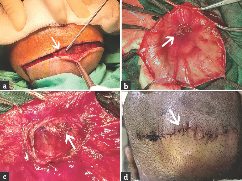
Figure 4.

Intraoperative images (white arrow): (a) Dissection of the flap along the marked skin margins, (b) exposure of the sac and the herniated nonviable cerebellar tissue, (c) double-layer closure of the dural defect, (d) skin closure 1 week postoperatively
