Figure 5.

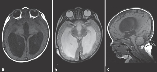
(a) Magnetic resonance imaging – T1, (b) magnetic resonance imaging – T2 axial, and (c) magnetic resonance imaging – T1 sagittal view 3 months postoperatively showing no encephalocele and resolved tonsillar herniation

(a) Magnetic resonance imaging – T1, (b) magnetic resonance imaging – T2 axial, and (c) magnetic resonance imaging – T1 sagittal view 3 months postoperatively showing no encephalocele and resolved tonsillar herniation