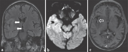
Figure 1.

A 40-year-old male patient with herpes simplex virus encephalitis presented with altered sensorium. Fluid-attenuated inversion recovery coronal image (a) shows hyperintensity in the right perisylvian and temporal regions (arrows). Diffusion-weighted imaging image (b) shows restricted diffusion in the right temporal region (arrowhead). Postcontrast T1-weighted images (c) shows mild enhancement in the right temporal cortex (open arrow)
