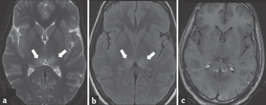
Figure 3.

A 15-year-old female patient with Japanese encephalitis presented with bilateral visual disturbances. T2W (a), T2 fluid-attenuated inversion recovery (b) images shows hyperintensity in bilateral posterior thalami (arrows). Postcontrast T1W image (c) shows no contrast enhancement
