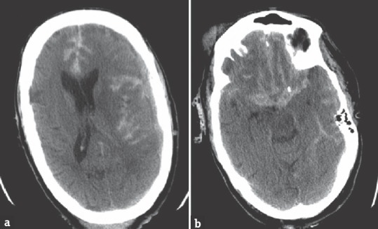
Figure 1.

Preoperative computed tomography scan demonstrates: (a) Frontotemporal subdural hemorrhage along the falx cerebri and midline shift compressing the left cerebral hemisphere; (b) diffuse subarachnoid hemorrhage extending from the suprasellar cistern
