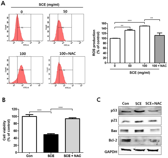
Figure 5.
Apoptotic effects of SCE are mediated in a ROS-dependent manner. (A) HCT116 cells were treated with the indicated concentrations of SCE, or 100 mg/ml SCE supplemented with NAC (5 mM) for 24 h. The production of ROS was estimated by fluorescence-activated cell sorting analysis with DCFDA staining. The fluorescence was analyzed by excitation/emission wavelength at 488/525 nm (indicated with PE-A). The representative figures are shown and DCFDA-positive cells (P2) were calculated as a percentage of the control (presented as the mean ± SEM). **P<0.01 and ***P<0.001 compared with the control. (B) The HCT116 cells were treated with SCE (100 µg/ml) with or without NAC (5 mM). The viabilities of the cells were measured by MTT assay and shown as a percentage of the control. ***P<0.001 compared with the control. (C) The total proteins were extracted from HCT116 cells treated with SCE (100 µg/ml) with or without NAC (5 mM). The expression levels of p53, p21, Bax, Bcl-2 and Mdm2 were examined by western blot analysis. The expression of GAPDH was used as an internal control. SCE, Sorbus commixta water extract; Bcl-2, B-cell lymphoma-2; Bax, Bcl-2-associated X protein; DCFDA, 2′,7′-dichlorodihydrofluorescein diacetate; NAC, N-acetyl cysteine.

