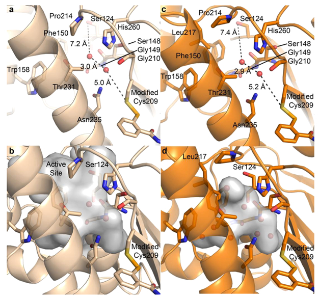
Figure 8.
Active site region differs in each structure. Atom color assignment is consistent with Figure 3, and water molecules are depicted by red spheres. (a) Residues that line the region of intrigue present in the azido ebselen structure are shown. The path from the Ser124 to the modified Cys209 is filled by two water molecules with distances shown. (b) Void volume depicted through a surface rendering of the cavity pocket found in the azido structure. (c) The void region observed in the adamantyl structure is lined with similar residues found in the azide structure, with the addition of Leu217. Again, two water molecules span the distance from Ser124 and Cys209, with distances nearly identical to a. (d) Void volume depicted through a surface rendering of the cavity pocket found in the adamantyl structure.

