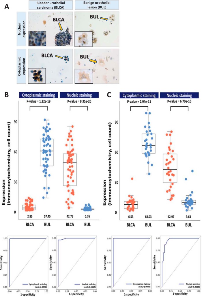
Fig. 5.
Results of for immunocytochemical staining of bladder urothelial carcinoma cells (BLCA, A) and benign urothelial cells (BUL, A) for AHNAK, and boxplot and beeswarm plot with the area under the receiver operating characteristic (ROC) curve (AUC) of individual immunocytochemical staining in two validation cohorts (B&C). A, Positive immunocytochemical staining for AHNAK in bladder urothelial carcinoma cells (left upper: nuclear expression and left lower: cytoplasmic expression. ×40, inset ×1000). Positive immunocytochemical staining for AHNAK in benign urothelial cells (right upper: nuclear expression and right lower: cytoplasmic expression. ×40, inset ×1000). (B and C) Average protein expression values are indicated by lines, and the median and quartile values of cells with AHNAK immunocytochemical staining are represented by boxes. p values obtained by nonparametric Wilcoxon test are shown on top of each bar graph. The AUC of AHNAK immunocytochemistry is shown to distinguish between benign urothelial lesion and bladder urothelial carcinoma.

