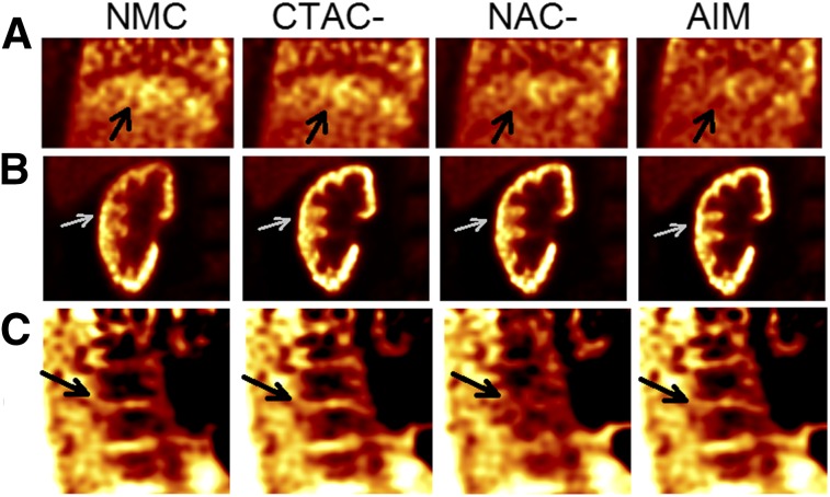
FIGURE 6.
Final reconstruction examples of 18F-FPDTBZ studies with different correction methods. (A) Coronal liver–lung region for study 1. Arrows point to lung–liver border. (B) Coronal right-kidney region for study 2. Arrows point to right-kidney cortex. (C) Sagittal spine region for study 1. Arrows point to bone marrow gap.

