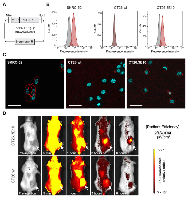
Figure 5. In vitro and in vivo evaluation of the new murine colorectal carcinoma model CT26.3E10 transfected with the human antigen CAIX.
(A) Cloning scheme of human CAIX in pcDNA3.1(+). (B) FACS analysis for expression of human CAIX on SKRC-52, CT26.wt and transfected CT26.3E10 cells. Staining was performed with a human anti-CAIX specific antibody and the corresponding signal was amplified with an anti-human AlexaFluor488 secondary antibody. (C) Confocal microscopy analysis on living SKRC-52, CT26.wt and CT26.3E10 cells after exposure to acetazolamide labeled with AlexaFluor594 (AAZ-AlexaFluor594; 5) (120 nM) at 1 hour incubation time. The conjugate is mainly found at the cell surface in CT26.3E10 and in the positive control SKRC-52. No cell surface binding can be detected for the negative control CT26.wt. Red = AAZ-AlexaFluor594 staining; Blue = Hoechst 33342 staining; Scale bar = 100 μm. (D) Evaluation of the in vivo targeting performance of AAZ-IRdye680RD (4) in immunocompetent BALB/c mice bearing CAIX transfected CT26.3E10 tumors by near-infrared fluorescence imaging. A selective tumor uptake at early time points (3 and 6 hours) was observed, in comparison to the biodistribution of the molecule in CAIX-negative CT26.wt tumor bearing mice.

