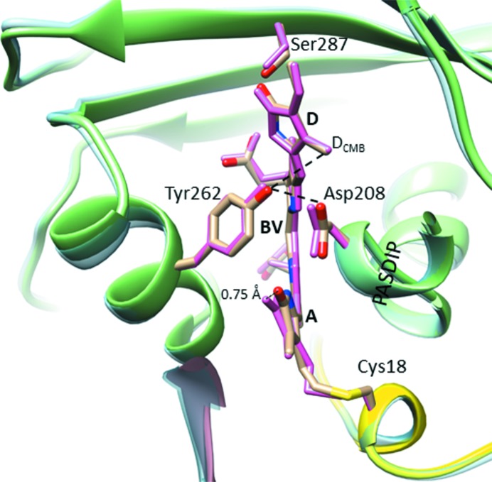
Figure 7.
Superposition of the T289H mutant SaBphP1-PCM structures determined at cryo (PAS, yellow; GAF, green; PHY, not shown) and at room temperatures (light-blue ribbon). The chromophore binding site is enlarged and important residues, the PASDIP consensus sequence and the BV chromophore are marked. Dark yellow residues from cryo structure; hot pink from the room-temperature structure.

