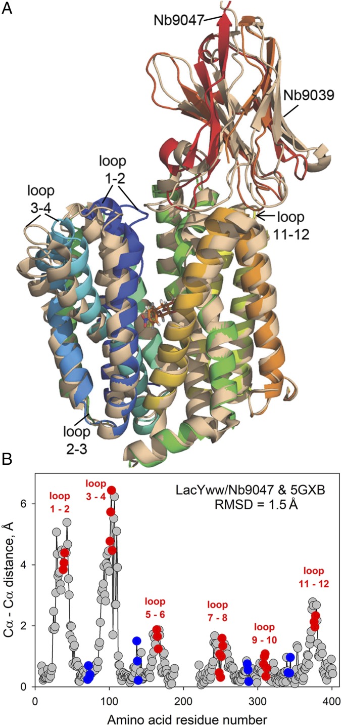
Fig. 2.
Structural alignment of two Nb-bound LacYWW complexes reveals a partial closure on the periplasmic side associated with ligand-binding, suggesting two states on the transport pathway. (A) Cartoon demonstrating two superimposed structures (with and without bound NPG) aligned by Cα atoms: LacYWW/NPG/Nb9047 (rainbow-colored as in Fig. 1A) and apo-LacYWW/Nb9039 (wheat-colored, PDB ID code 5GXB). Nbs are attached to the periplasmic side of LacYWW. (B) Differences in positions of the main-chain Cα atoms of LacYWW between the two aligned structures based on residues 9–188 and 221–401 (Nbs and NPG removed). Amino acids in loop 6–7 and in N- and C-ends are omitted (rmsd = 1.5 Å). Red and blue dots represent residues in periplasmic and cytoplasmic loops, respectively.

