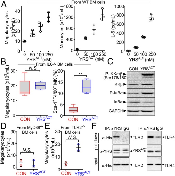
Fig. 7.
Effect of YRSACT depends on the TLR/MyD88 signaling pathway. (A) BM cells from three WT mice were pooled and split into aliquots for treatment with different YRSACT concentrations for 3 d. Cells were analyzed for MK (Left) and monocyte (Middle) count; additionally, culture supernatants were collected to measure IL-6 by ELISA (Right). Each experiment was performed with technical triplicates. (B) BM cells from IL-6−/− mice (n = 4) were isolated and treated with YRSACT for 3 d for evaluation by flow cytometry. (C) Normal hPBMCs were treated with YRSACT or PBS (CON) for 20 min and then lysed and analyzed by Western blotting to evaluate NF-κB activation. (D) BM cells from three MyD88−/− mice were pooled and treated with 100 nM YRSACT or PBS (CON) for 3 d before analysis by flow cytometry (n = 2 with technical triplicates). (E) BM cells prepared from three TLR2−/− mice were treated with YRSACT or PBS (CON) for 3 d before analysis by flow cytometry. (F) YRSACT was incubated with His-tagged recombinant TLR2 or TLR4 and then mixed with anti-YRS polyclonal antibody or control IgG; the precipitate was then analyzed by immunoblotting with an anti-His tag antibody. IP, immunoprecipitation. Data are shown as dot plots with mean ± SD in A, D, and E, or as 25th to 75th percentile bars with median and min to max whiskers in B; *P < 0.01 determined by Mann–Whitney two-tailed unpaired t test. N.S., not significant.

