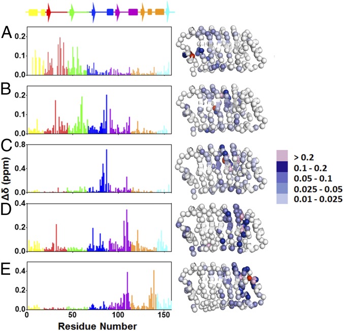
Fig. 2.
Chemical shift perturbations of the cavity-containing variants relative to WT pp32. (A) I7A, (B) L60A, (C) L83A, (D) L109A, and (E) L139A variants. The structures, based on the crystal structure of pp32 (19), depict the backbone nitrogen atoms as spheres. They were rendered using Pymol (23). The backbone nitrogen at the site of the mutation is colored in red. Residues are colored for the amplitude of the chemical shift as indicated in the scale to the Right of the structures.

