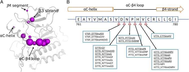
Fig. 1.
EGFR insertion mutations. (A) Structural location of insertion mutations in the EGFR kinase domain crystal structure (PDB ID: 2GS6). The size of the magenta sphere is proportional to the log of the number of patient samples containing the insertion mutation at the particular residue position. (B) Sequence location of insertion mutations within the EGFR C–4 loop. The nomenclature of insertion mutations follows the guidelines established previously (42). For example, V769_D770insASV indicates an Ala–Ser–Val insertion between residues V769 and D770, and D770GY indicates a complex insertion mutation in which D770 is replaced by a Gly–Tyr sequence. Insertion mutations characterized in this study are underlined. The complete list of patient-derived insertion mutations in EGFR can be found in SI Appendix, Dataset S1.

