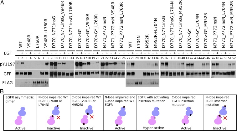
Fig. 3.
Dimerization dependency of EGFR insertion mutations. (A) Western blot analysis of the N-lobe dimerization-deficient mutation (L760R or L704N) and the C-lobe dimerization-deficient mutation (V948R or M952R) on Y1197 autophosphorylation for WT and mutant (D770_N771insG, N771_P772insN, and D770GY) EGFR. All of the constructs used are tagged with GFP except for L760R and L704N, which are tagged with FLAG. Cotransfection of V948R and L760R is indicated by V948RL760R. Co-transfection of M952R and L704N is indicated by M952RL704N. Eight percent SDS/PAGE is used to resolve the cell lysate sample. Y1197 phosphorylation of V948RL760R and M952RL704N is also analyzed by 5% SDS/PAGE to separate GFP- and FLAG-tagged constructs (SI Appendix, Fig. S1 A and B). Densitometry analysis of three to five independent experiments is shown in SI Appendix, Fig. S2). (B) A schematic plot summarizing the observed differences in WT and mutant EGFR in the different dimerization-deficient mutant backgrounds.

