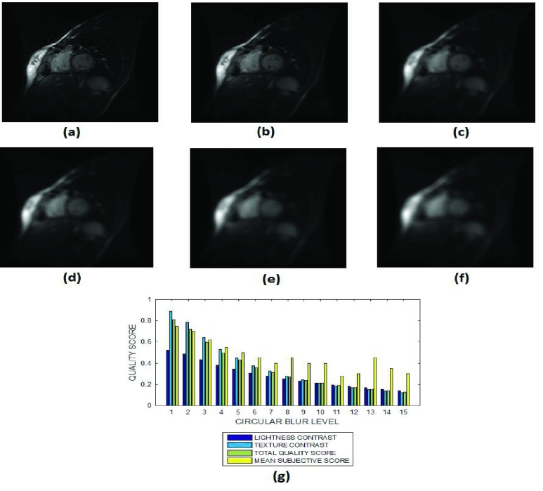
FIGURE 8.
(a) A short axis cardiac MRI slice and its degraded versions at circular blur levels (b) 3 voxels, (c) 7 voxels, (d) 9 voxels, (e) 12 voxels and (g) 15 voxels, (h) variation of the lightness contrast, texture contrast, total quality scores and the mean subjective scores with blur levels increasing from 1 to 15.

