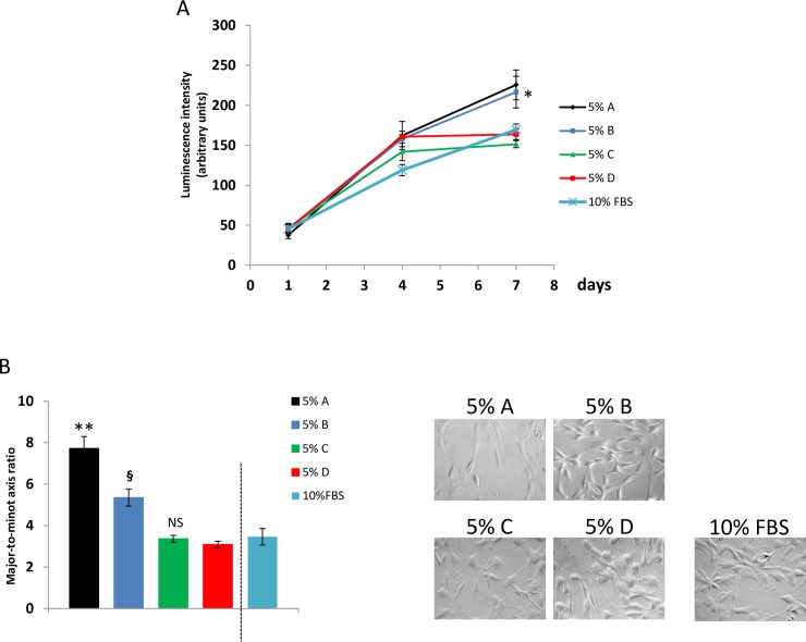
Fig 1.
Impact of medium additives A, B, C and D on proliferation rate of ASC. (A) Cell growth test based on luminescence generated by living and proliferating cells. Results show that in presence of 5% medium additive A (recalcified PRP) and B (recalcified PPP) cell proliferation rate is higher, when compared to additives C (freeze and thaw, plasma depleted platelets) and D (freeze and thaw, PRP). (B) Mean values of major-to-minor axis ratios and representative images of ASC expanded in presence of the different additives. ASC expanded in presence of medium additives A and B were more elongated than ASC expanded in C and D. ASC expanded with additive A were more elongated than ASC cultured with additive B. Values of luminescence and of major-to-minor axis ratios obtained from cells expanded in presence of 10% FBS were shown as control condition. Considering such results, ASC expanded in presence of medium additive A showed higher proliferation rate when compared to additive B. When compared to medium additives A and B, ancillary products C and D weakly stimulated cell proliferation. ANOVA for independent samples was performed as statistical analysis (p<0.001) and Tukey’s honestly different significance with Bonferroni’s correction was chosen as post hoc test to compare the effects of the different medium additives.*, p<0.05 vs C and D; **, p<0.01 vs B, C and D; §, p<0.05 vs C and D; NS vs D.

