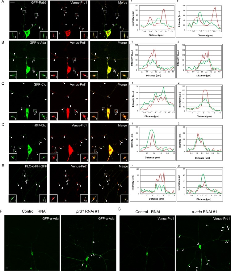
Fig 2. Prd1 colocalizes with α-Ada and clathrin in dendrites.
(A) Distribution of Venus-Prd1 and GFP-Rab5 in ddaC neurons. Venus-Prd1 primarily localized adjacent to GFP-Rab5 (open arrowheads) (insets) and occasionally colocalized with GFP-Rab5 (arrowheads) in the dendrites. (B) Distribution of GFP-α-Ada and Venus-Prd1 in ddaC neurons. Venus-Prd1 colocalized with GFP-α-Ada in the dendrites (arrowheads). (C) Distribution of Venus-Prd1 and GFP-Clc in ddaC neurons. Venus-Prd1 colocalized with GFP-Clc in the dendrites (arrowheads). (D) Distribution of Venus-Prd1 and mRFP-Chc in ddaC neurons. Venus-Prd1 colocalized with mRFP-Chc in the dendrites (arrowheads). (E) Distribution of Venus-Prd1 and PLC-δ-PH-GFP in ddaC neurons. Venus-Prd1 colocalized with PLC-δ-PH-GFP in the dendrites (arrowheads). Line profiles show the arbitrary fluorescence intensity along the white lines. (F) Distribution of GFP-α-Ada in control and prd1 RNAi ddaC neurons. (G) Distribution of Venus-Prd1 in control and α-ada RNAi ddaC neurons. Dorsal is up in all images. Scale bars in (A) and (F) represent 10 μm. The individual numerical values for panels A, B, C, D, and E can be found in S1 Data. The genotypes can be found in S1 Text. α-Ada, α-Adaptin; a.u., arbitrary unit; Chc, Clathrin heavy chain; Clc, Clathrin light chain; GFP, green fluorescent protein; mRFP, monomeric red fluorescent protein; PLC-δ-PH, phospholipase C-δ-pleckstrin homology; Prd1, pruning defect 1; Rab5, Rabaptin-5; RNAi, RNA interference.

