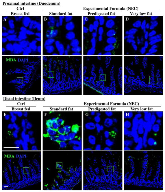
Figure 6. The effect of fat composition on the accumulation of oxidized lipids in ileal enterocytes of mice induced to develop NEC.
A–H: Immunofluorescence images of Malondialdehyde (MDA-green) and DAPI stained (nuclei, blue) from control and NEC mice proximal (duodenum, A–D) and distal (ileum, E–H) small intestine 10μm, cryo-sections) (Ctrl, Control-Breast fed or experimental NEC treatments with hypoxia and formula feeding). PDF, pre-digested fat, scale bar =10μm.

